SVT vs sinus tachycardia: how to tell them apart on an ECG
A fast, narrow, regular rhythm usually comes down to one question: is the heart answering a call, or is it stuck in a loop?
Sinus tachycardia is the normal heartbeat running faster because the body needs it to: exercise, pain, fever, blood loss, low oxygen. It speeds up and slows down gradually, and there is a P wave in front of every QRS.1 Most paroxysmal SVT is an impulse caught in a loop through or beside the AV node. It switches on and off suddenly, usually runs at 150 to 220 beats a minute, and its P waves are hidden or sit just after the QRS.2 Because the loop runs through the AV node, a vagal maneuver or adenosine can break it. Sinus tachycardia only slows for a moment, so the answer there is to treat the cause.1,2
| Sinus tachycardia | SVT (AVNRT, AVRT) | |
|---|---|---|
| Why it's fast | The body is asking for more output | A re-entry loop, whatever the body needs |
| Start and stop | Gradual | Sudden on, sudden off |
| Usual rate | Above 100, matching the need | 150 to 220 |
| Beat to beat | Drifts a little with breathing and activity | Fixed, regular as a metronome |
| P waves | Upright in I and II, one before each QRS | Hidden in the QRS, or just after it |
| Vagal maneuver or adenosine | Slows for a moment, then speeds back up | Often stops it at once |
Answering: sinus tachycardia, 140 a minute
Looping: AVNRT, 170 a minute
What do both rhythms have in common?
Every normal beat follows the same route. It starts in the sinoatrial (SA) node, high in the right atrium, and spreads across both atria. The atrioventricular (AV) node then holds it for about a tenth of a second, giving the atria time to finish emptying into the ventricles.2 From there it races down the His bundle, the bundle branches and the Purkinje fibers into both ventricles at once.
That fast lane is why a normal QRS is narrow, under 120 ms. A narrow-complex tachycardia tells you the ventricles are still being activated through it, so the rhythm starts at or above the AV node.2 Sinus tachycardia and SVT both qualify, and that is why they can look so alike.
What is sinus tachycardia?
In sinus tachycardia nothing about the route changes. The SA node simply fires faster than 100 times a minute, because the nervous system and circulating catecholamines are asking it to.1
Sometimes the reason is ordinary: exercise, stress, pain or anxiety. Sometimes it's serious: fever and sepsis, blood loss, dehydration, anemia, low oxygen, a pulmonary embolism, an overactive thyroid, or stimulants such as cocaine and amphetamines.1 A fast heart rate at rest can be one of the earliest signs that something is wrong, so it deserves an explanation rather than a drug.1
On the ECG it looks like normal sinus rhythm, only faster. Each QRS has a P wave in front of it, upright in leads I and II and negative in aVR.1 The rate climbs and settles gradually as the cause comes and goes. At very fast rates the P wave starts to crowd into the T wave of the beat before, and that is when sinus tachycardia gets hard to tell from SVT.
140 a minute. The gap between each T wave and the next P wave is closing. The P wave is still there, in front of every QRS.
What is SVT?
Strictly, supraventricular tachycardia is an umbrella term for any fast rhythm that starts at or above the AV node, and it takes in sinus tachycardia, atrial tachycardia and atrial flutter.2,3 In everyday use, and on this page, "SVT" means paroxysmal SVT: the sudden, regular re-entry rhythms. The two common ones are AVNRT and AVRT.2
Re-entry means an impulse caught in a circuit, going around and around and firing the ventricles on every lap.
- AVNRT (AV nodal re-entrant tachycardia) is the most common type in adults. The AV node has two inputs, a fast pathway and a slow pathway, and the loop runs down one and back up the other, through the tissue in and around the AV node.4
- AVRT (AV re-entrant tachycardia) needs an extra connection between the atria and the ventricles, an accessory pathway, as in Wolff-Parkinson-White syndrome. In the usual, orthodromic form the impulse goes down through the AV node and back up the accessory pathway, so the QRS stays narrow.5
AVNRT loops through and around the AV node
AVRT loops down the AV node and back up an accessory pathway
Because the loop doesn't follow the body's needs, SVT tends to start suddenly, often with a premature beat, and stop just as abruptly. It usually runs at 150 to 220 beats a minute and is very regular.2 People often describe a sudden pounding that switches on and off. Caffeine, alcohol, stimulants and stress can be triggers.2
How do they look side by side on an ECG?
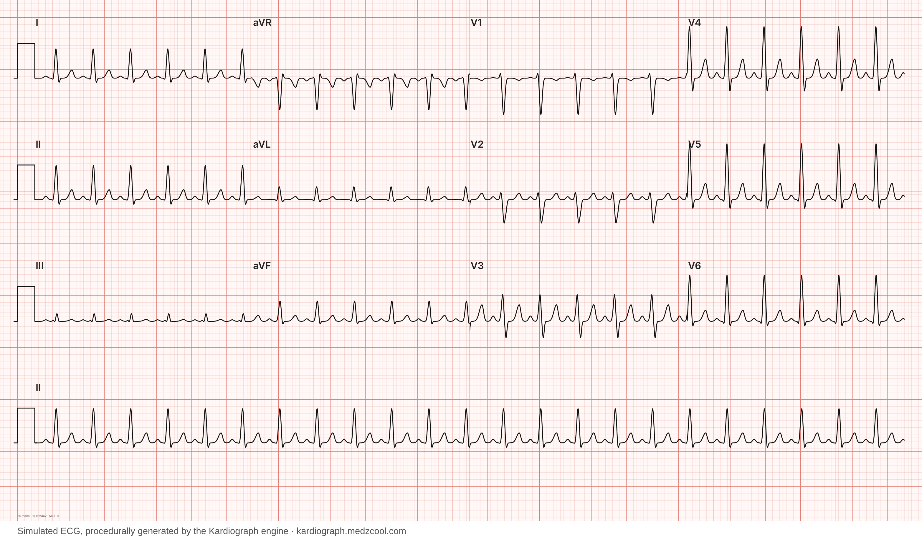
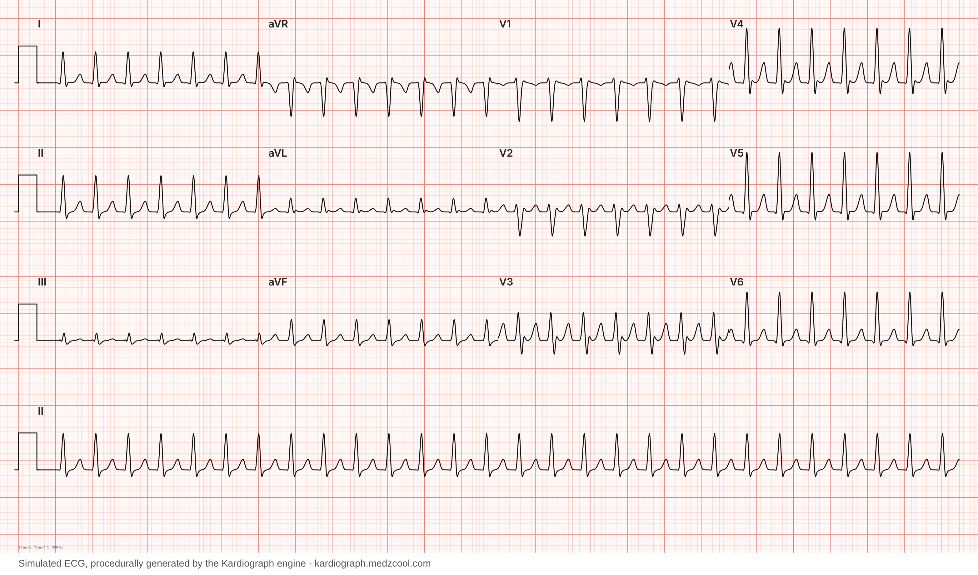
Look for the P waves first, in lead II and V1.
- Sinus tachycardia (140 a minute): a P wave before every QRS, upright in I and II, with a normal PR interval. The space between the T wave and the next P wave is short, but it's there.
- AVNRT (170 a minute): no P wave in front of the QRS. The atria fire backward, from the AV node upward, at almost the same moment as the ventricles, so the P wave is buried in the QRS or sits just after it. It can show up as a small pseudo-r′ at the end of the QRS in V1, or a pseudo-S wave in II, III and aVF.4
Then look at the rate and how it behaves. A rate above about 150 at rest makes SVT more likely, but rate alone can't settle it. A record of the start or end of the episode helps the most: a sudden switch points to SVT, a gradual ramp to sinus tachycardia.2
Why do vagal maneuvers and adenosine stop SVT but not sinus tachycardia?
Both slow conduction through the AV node for a moment. If the loop runs through the AV node, as it does in AVNRT and orthodromic AVRT, a brief block breaks it, and sinus rhythm takes over again.2,3 Sinus tachycardia has no loop to break. The rate dips for a few seconds and then comes back, because the reason it was fast is still there.
For a patient with a regular, narrow-complex tachycardia who is stable, the guidelines start with vagal maneuvers, done lying down.2,3,6 The modified Valsalva maneuver tested in the REVERT trial works best: a 15-second strain while sitting semi-upright, then lying flat straight away with the legs lifted. In that trial 43% of patients went back to sinus rhythm, against 17% with a standard Valsalva.7 If that fails, adenosine is the first-line drug. It is given by rapid IV push with the patient on a monitor and the ECG recording, which also helps diagnose the rhythm if it doesn't stop.2,3
A patient who is unstable, with low blood pressure, chest pain, low oxygen, confusion or other signs of shock, gets synchronized cardioversion instead.2,3
Two safety rules matter here:
- Never slow a sinus tachycardia that is compensating for blood loss, sepsis, dehydration or low oxygen. The fast rate is keeping the patient's output up. Treat the cause.1
- This page covers regular, narrow-complex tachycardia only. An irregular or wide-complex fast rhythm may be atrial fibrillation conducting down an accessory pathway (pre-excited AF), where drugs that block the AV node can let the ventricles race and degenerate into ventricular fibrillation.2,5,6
This page is for learning to recognize these rhythms. It is not a treatment protocol, and it leaves out drug doses.
What can fool you?
- Atrial flutter with 2:1 conduction. The atria flutter at about 300 a minute and every second wave gets through, giving a regular, narrow rhythm at about 150 that can pass for either. Look for the sawtooth: continuous negative flutter waves in II, III and aVF.8 One of them often hides in the QRS or the T wave. Adenosine doesn't stop flutter, because the AV node isn't part of its loop,2 but the brief block can slow the ventricles enough to unmask the flutter waves.9
- Very fast sinus tachycardia. In a young adult exercising, or a patient who is very sick, sinus rates can climb into the 150s and above, and the P wave can sit on top of the T wave before it. The clues are the gradual change in rate and a cause you can find.
- Focal atrial tachycardia. A single spot in the atrium fires fast instead of the SA node. There is a P wave before each QRS, but it often has a different shape or axis from a sinus P wave, and adenosine often doesn't stop it, because the AV node isn't part of the problem.2
References
- Henning A, Krawiec C. Sinus Tachycardia. In: StatPearls. Treasure Island (FL): StatPearls Publishing; updated 2023 Mar 5. www.ncbi.nlm.nih.gov/books/NBK553128/
- Patti L, Horenstein MS, Ashurst JV. Supraventricular Tachycardia. In: StatPearls. Treasure Island (FL): StatPearls Publishing; updated 2025 Feb 18. www.ncbi.nlm.nih.gov/books/NBK441972/
- Page RL, Joglar JA, Caldwell MA, et al. 2015 ACC/AHA/HRS Guideline for the Management of Adult Patients With Supraventricular Tachycardia. Circulation. 2016;133(14):e506-e574. doi:10.1161/CIR.0000000000000311 · PubMed 26399663
- Hafeez Y, Ahmed I. Atrioventricular Nodal Reentry Tachycardia. In: StatPearls. Treasure Island (FL): StatPearls Publishing; updated 2026 Mar 25. www.ncbi.nlm.nih.gov/books/NBK499936/
- Chhabra L, Goyal A, Benham MD. Wolff-Parkinson-White Syndrome. In: StatPearls. Treasure Island (FL): StatPearls Publishing; updated 2023 Aug 7. www.ncbi.nlm.nih.gov/books/NBK554437/
- Brugada J, Katritsis DG, Arbelo E, et al. 2019 ESC Guidelines for the management of patients with supraventricular tachycardia. Eur Heart J. 2020;41(5):655-720. doi:10.1093/eurheartj/ehz467 · PubMed 31504425
- Appelboam A, Reuben A, Mann C, et al. Postural modification to the standard Valsalva manoeuvre for emergency treatment of supraventricular tachycardias (REVERT): a randomised controlled trial. Lancet. 2015;386(10005):1747-1753. doi:10.1016/S0140-6736(15)61485-4 · PubMed 26314489
- Rodriguez Ziccardi M, Goyal A, Maani CV. Atrial Flutter. In: StatPearls. Treasure Island (FL): StatPearls Publishing; updated 2022 Dec 8. www.ncbi.nlm.nih.gov/books/NBK540985/
- Patel P, Goyal A. Antiarrhythmic Medications. In: StatPearls. Treasure Island (FL): StatPearls Publishing; updated 2024 Feb 28. www.ncbi.nlm.nih.gov/books/NBK482322/